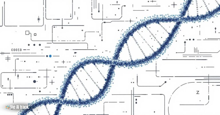
Researchers at the University of Missouri have developed a cutting-edge AI tool that reconstructs the 3D chromosome structure in single cells, dramatically improving accuracy in gene architecture modeling. This innovation enables deeper understanding of gene expression regulation, disease mechanisms, and cell-specific genomic variation.
New AI Tool Predicts 3D Chromosome Structures – Key Points
Innovators:
Graduate student Yanli Wang and Professor Jianlin “Jack” Cheng from the University of Missouri’s College of Engineering led the development of the AI tool. Their work addresses a critical gap in analyzing chromosome structures at single-cell resolution.
Technological Advancement:
The AI model uses SO(3)-equivariant graph neural networks (GNNs)—a form of machine learning well-suited for processing 3D spatial structures that are rotationally invariant. This makes it possible to accurately interpret chromosomal data regardless of orientation in the nucleus.
Enhanced Accuracy:
The new model outperforms previous state-of-the-art methods by over two-fold in accuracy when reconstructing 3D chromosome structures from noisy, sparse single-cell Hi-C data. It models chromatin contacts as spatial graphs and can predict structure even with incomplete data.
Significance of Chromosome Folding:
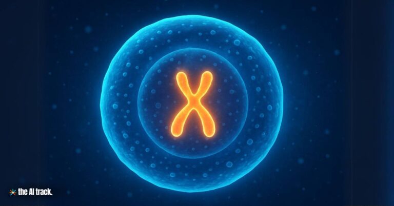
Each cell contains approximately six feet of DNA that must fold correctly to fit in the nucleus and regulate gene expression. Improper folding or chromatin misorganization is linked to diseases such as cancer and developmental disorders. This tool helps detect these misfolds at the individual-cell level.
Limitations of Previous Methods:
Prior methods relied on averaged data from millions of cells, obscuring crucial variations between individual cells. This model enables a cell-by-cell view of chromosomal folding, capturing heterogeneity that affects gene activity and cell function.
Biological Implications:
The ability to map 3D chromosome structure in single cells opens new directions in developmental biology, personalized medicine, and cancer diagnostics. Subtle structural variations can now be linked to phenotypic diversity and pathological changes.
Accessibility:
The software has been made freely available to researchers globally, democratizing access and accelerating genomics and biomedical research across cell types and disease models.
Future Developments:
The team aims to expand the AI tool to reconstruct full 3D genome architectures at high resolution, offering the most detailed blueprint of genetic organization to date. This could transform precision medicine and diagnostic technologies.
Publication:
The study, titled “Reconstructing 3D chromosome structures from single-cell Hi-C data with SO(3)-equivariant graph neural networks,” was published in NAR Genomics and Bioinformatics on March 22, 2025. DOI: 10.1093/nargab/lqaf027
Why This Matters:
This AI model represents a major leap in genomic modeling. By resolving individual cell chromosomal structures with high accuracy, researchers can now dissect how gene expression is physically controlled within the nucleus—providing deeper insights into disease mechanisms, developmental biology, and cell-specific functions. The implications for genomic medicine, biotech, and AI-assisted research are profound.
A curated collection of the most significant recent AI breakthroughs in biology, advancing our understanding of genetics and ecosystems.
Read a comprehensive monthly roundup of the latest AI news!